Back Of Neck Nerves Anatomy - Nerve roots exit the spinal cord in the neck and provide control and sensation to different parts of the body.. 2 (c1 does not have a dermatome.) Nerves in the neck, medically referred to as the cervical spine, help transmit information along the pathways of the central and peripheral nervous system, including sensory and motor skills processes. The cervical spine protects the nerves connecting to. C1, c2, and c3 (the first three cervical nerves) help control the head and neck, including movements forward, backward, and to the sides. In the cervical spine, the spinal cord connects to the brain at the base of the skull.
Amongst these components is the ansa cervicalis: The cervical spine is the top part of the spine. 2 (c1 does not have a dermatome.) The peripheral nerves include both motor nerves and sensory nerves: Peripheral nerves comprise the peripheral nervous system.
Innervation of the heart: Sympathetic and parasympathetic ... from thumbor.kenhub.com 1 the c2 dermatome handles sensation for the upper part of the head, and the c3 dermatome covers the side of the face and back of the head. C1, c2, and c3 (the first three cervical nerves) help control the head and neck, including movements forward, backward, and to the sides. The cervical spine protects the nerves connecting to. Eight spinal nerves branch off from the spinal cord in the neck to form a network of nerves called the cervical plexus. Your neck is like no other part of the vertebral spinal column and enables your head and neck a wide range of motion. Nerves in the neck, medically referred to as the cervical spine, help transmit information along the pathways of the central and peripheral nervous system, including sensory and motor skills processes. The posterior root, located in back, carries sensory signals from the body back to the brain. Nov 02, 2020 · in the neck, the spinal cord passes through the vertebral foramen of the cervical vertebrae, which surround and protect its delicate nervous tissue.
These nerves are made up of nerve fibers, called sensory fibers (mechanoreceptor fibers sense body movement and pressure against the body, and.
More images for back of neck nerves anatomy » The numerous motor components of the cervical plexus mainly innervate the muscles of the neck and upper back. Nov 02, 2020 · in the neck, the spinal cord passes through the vertebral foramen of the cervical vertebrae, which surround and protect its delicate nervous tissue. Eight spinal nerves branch off from the spinal cord in the neck to form a network of nerves called the cervical plexus. May 06, 2015 · nerves within the cervical spine: The cervical column is comprised of 7 bones (c1 to c7) uniquely shaped to protect the spinal cord that descend from the base of your skull and the spinal nerves or root that exit the spine between each set of bones. The peripheral nerves include both motor nerves and sensory nerves: Amongst these components is the ansa cervicalis: C1, c2, and c3 (the first three cervical nerves) help control the head and neck, including movements forward, backward, and to the sides. Your neck is like no other part of the vertebral spinal column and enables your head and neck a wide range of motion. There are 8 spinal nerves that originate from the cervical spine. From there, the spinal nerve branches into a network of nerves. Sensory nerves are nerves that receive sensory stimuli, telling us how something feels—whether it is hot, cold, or painful.
The cervical column is comprised of 7 bones (c1 to c7) uniquely shaped to protect the spinal cord that descend from the base of your skull and the spinal nerves or root that exit the spine between each set of bones. The cervical spine is the top part of the spine. Amongst these components is the ansa cervicalis: Each nerve provides sensation to a specific area of the body called a dermatome. Nerves in the neck, medically referred to as the cervical spine, help transmit information along the pathways of the central and peripheral nervous system, including sensory and motor skills processes.
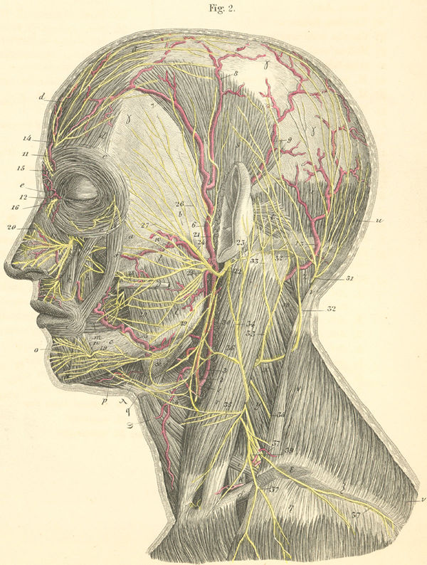
Superior view of the nerves of the head and neck (left side) from www.anatomyatlases.org 2 (c1 does not have a dermatome.) Amongst these components is the ansa cervicalis: Nerves in the neck, medically referred to as the cervical spine, help transmit information along the pathways of the central and peripheral nervous system, including sensory and motor skills processes. 1 the c2 dermatome handles sensation for the upper part of the head, and the c3 dermatome covers the side of the face and back of the head. It is formed at the posterior border of the anterior scalene muscle by fibres from c3, 4 and 5 nerve roots. The cervical spine is the top part of the spine. C1, c2, and c3 (the first three cervical nerves) help control the head and neck, including movements forward, backward, and to the sides. More images for back of neck nerves anatomy »
The peripheral nerves include both motor nerves and sensory nerves:
It runs from the neck to the upper back. The cervical spine is the top part of the spine. Peripheral nerves comprise the peripheral nervous system. Nerves in the neck, medically referred to as the cervical spine, help transmit information along the pathways of the central and peripheral nervous system, including sensory and motor skills processes. Eight spinal nerves branch off from the spinal cord in the neck to form a network of nerves called the cervical plexus. These nerves are made up of nerve fibers, called sensory fibers (mechanoreceptor fibers sense body movement and pressure against the body, and. It consists of seven vertebrae. The majority of these nerves control the functions of the upper extremities and allow you to feel your arms, shoulder, and back of your head. The posterior root, located in back, carries sensory signals from the body back to the brain. It is formed at the posterior border of the anterior scalene muscle by fibres from c3, 4 and 5 nerve roots. In the cervical spine, the spinal cord connects to the brain at the base of the skull. C1, c2, and c3 (the first three cervical nerves) help control the head and neck, including movements forward, backward, and to the sides. Dec 04, 2018 · anatomy of the neck spinal cord.
In the cervical spine, the spinal cord connects to the brain at the base of the skull. A nerve that exits the lower back has peripheral branches that extend all the way down to the toes. Nerves in the neck, medically referred to as the cervical spine, help transmit information along the pathways of the central and peripheral nervous system, including sensory and motor skills processes. Your neck is like no other part of the vertebral spinal column and enables your head and neck a wide range of motion. Each nerve provides sensation to a specific area of the body called a dermatome.
Main nerves of the head and neck (preview) - Human Anatomy ... from i.ytimg.com The peripheral nerves include both motor nerves and sensory nerves: A nerve that exits the lower back has peripheral branches that extend all the way down to the toes. The numerous motor components of the cervical plexus mainly innervate the muscles of the neck and upper back. May 06, 2015 · nerves within the cervical spine: Dec 04, 2018 · anatomy of the neck spinal cord. These 2 nerve roots branch directly from the spinal cord and merge to form the spinal nerve as it runs through an opening between adjacent vertebrae, called the intervertebral foramen. The cervical column is comprised of 7 bones (c1 to c7) uniquely shaped to protect the spinal cord that descend from the base of your skull and the spinal nerves or root that exit the spine between each set of bones. There are 8 spinal nerves that originate from the cervical spine.
A nerve that exits the lower back has peripheral branches that extend all the way down to the toes.
It runs from the neck to the upper back. Nov 02, 2020 · in the neck, the spinal cord passes through the vertebral foramen of the cervical vertebrae, which surround and protect its delicate nervous tissue. Eight spinal nerves branch off from the spinal cord in the neck to form a network of nerves called the cervical plexus. The numerous motor components of the cervical plexus mainly innervate the muscles of the neck and upper back. There are 8 spinal nerves that originate from the cervical spine. Nerves in the neck, medically referred to as the cervical spine, help transmit information along the pathways of the central and peripheral nervous system, including sensory and motor skills processes. 1 the c2 dermatome handles sensation for the upper part of the head, and the c3 dermatome covers the side of the face and back of the head. Each nerve provides sensation to a specific area of the body called a dermatome. The cervical column is comprised of 7 bones (c1 to c7) uniquely shaped to protect the spinal cord that descend from the base of your skull and the spinal nerves or root that exit the spine between each set of bones. Amongst these components is the ansa cervicalis: Your neck is like no other part of the vertebral spinal column and enables your head and neck a wide range of motion. The cervical spine protects the nerves connecting to. May 06, 2015 · nerves within the cervical spine:
There are 8 spinal nerves that originate from the cervical spine back of neck anatomy. It is formed at the posterior border of the anterior scalene muscle by fibres from c3, 4 and 5 nerve roots.


0 Komentar